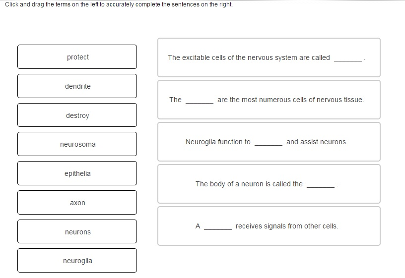
Match Each Label With the Medical Imaging Technique It Describes
31 Medical Imaging Technologies 14 Terms. Match each label with the medical imaging technique it describes.

Solved Match Each Label With The Medical Imaging Technique Chegg Com
We call these scans.

. Diagnostic imaging is non-invasive meaning medical professionals can look inside without surgery. Each kidney contains approximately 6-10 pyramids. A single barrage of x-rays passes through the body producing an image of interior structures on x-ray.
The upper limbs are held out to each side and the palms of the hands face forward as illustrated in Figure 141. Match each organ system with its functions. Using this standard position reduces confusion.
Match specific tooth views to specified tooth mouth windows. Mitochondria are cellular organelles more numerous in active cells. With these exams doctors can see how inner organs are functioning joints are moving and much more.
The brachial artery gives rise to the ulnar and radial arteries. These diagnostic imaging techniques are the work of radiologic technologists who use their powers to help save livesall without a cape. By imaging a patient in standard anatomical position a radiologist can build an X-Y-Z axis around the patient to apply body planes to the images.
However this activation Mo-99 has relatively low specific activity with a maximum of 74 GBqg depending on the neutron flux available in the reactor compared with 185 TBqg or. Radiographic visualization of blood vessels and blood flow by injecting radiopaque material through a. Experts are tested by Chegg as specialists in their subject area.
A section is a two-dimensional surface of a three-dimensional structure that has been cutModern medical imaging devices enable clinicians to obtain virtual sections of living bodies. Medical imaging refers to several different technologies that are used to view the human body in order to diagnose monitor or treat. An MRI is a diagnostic technique used to produce a detailed image of the bodies tissue and bones.
Medical imaging Techniques that are essential for diagnosing a wide range of disorders. Medical imaging is the technique of producing visual representations of areas inside the human body to diagnose medical problems and monitor treatment. Positron emission tomography PET is a medical imaging technique involving the use of so-called radiopharmaceuticals substances that emit radiation that is short-lived and therefore relatively safe to administer to the body.
Describe techniques for patient management while acquiring radiographic images including for patients with special needs. Medical imaging techniques such as sonography CT scans MRI scans or PET scans are one of the primary applications of body planes. It uses magnetic fields and radio waves to produce these images.
Although the first PET scanner was introduced in 1961 it took 15 more years before radiopharmaceuticals were combined with the technique and. Label the left and right pleural cavities and the mediastinum. The grandparent of all medical imaging techniques.
Conventional radiography X-rays pass through the body and expose on X- ray film producing a negative image called a roentgenogram. However in medical imaging the cost of Mo-99 itself is small relative to hospital costs. Terms in this set 24 Angiography.
It has had a. Mo-99 can also be made by bombarding Mo-98 with neutrons in a reactor. Who are the experts.
Computed tomography scanning CT or computerized axial tomography CAT X-rays pass. Positron emission tomography PET is a medical imaging technique involving the use of so-called radiopharmaceuticals substances that emit radiation that is short-lived and therefore relatively safe to administer to the body. We review their content and use your feedback to keep the quality high.
Is a medical imaging technique consisting of X-ray computed tomography where the X-rays are computed using cone beam technology. Brain Imaging Scanning Techniques Neuroimaging Below is a compilation of brain imaging neuroimaging or brain scanning techniques that have been utilized throughout history along with a brief description of how they work the associated advantages and disadvantages associated with each development. We first use a set of keywords describing medical imaging techniques such as MRI or ultrasound see AppendixB to match across the caption and reference text discarding images where a keyword does not appear.
Although the first PET scanner was introduced in 1961 it took 15 more years before radiopharmaceuticals were combined with the technique and. Where the value of m is selected such that i B m and θ jmns k 1 represents the probability that rater j observed label n given that the true label is s in region B mThe prior probability fT i s can be either a global prior or a voxelwise priorIn the case of this paper f T i s is a global prior that represents an a priori estimate of the fraction of voxels in the true. Chapter 22 Medical Imaging.
Describe medical imaging techniques in terms of their function and use in the diagnosis of diseases. Body sections and scans can be correctly interpreted however only if the viewer understands the plane along which the section was made. Terms in this set 14.
The human heart is comprised of four chambers. For example a scar in the anterior front carpal wrist. After filtering by keyword some images are still non-medical eg some are natural images of medical imaging equipment or graphs.
Match each description with medical imaging techniques. It does not matter how the body being described is oriented the terms are used as if it is in anatomical position. Medical imaging techniques have brought about tremendous progress in the field of medicine by virtually eliminating exploratory surgeries and.
Radiography x-rays Radiography Procedure. Match each label with the medical imaging technique it describes. Show transcribed image text.
The techniques and procedures used to create images of the human body.
Solved Click And Drag Each Label To The Appropriate Category Chegg Com
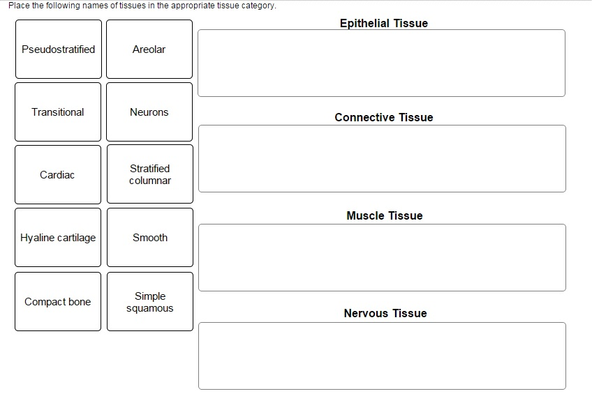
Solved Click And Drag Each Label To The Appropriate Category Chegg Com

The Core Penumbra Model Implications For Acute Stroke Treatment And Patient Selection In 2021 Baron 2021 European Journal Of Neurology Wiley Online Library

Sensors Free Full Text Deep Learning For Smart Healthcare Mdash A Survey On Brain Tumor Detection From Medical Imaging Html

Bir Publications

Anat2551hwsol6 Pdf 11 Award 10 00 Points Problems Adjust Credit For All Course Hero
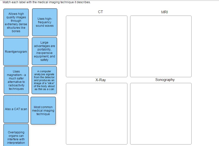
Match Each Label With The Medical Imaging Technique Chegg Com

Personalized Breast Cancer Treatments Using Artificial Intelligence In Radiomics And Pathomics Journal Of Medical Imaging And Radiation Sciences
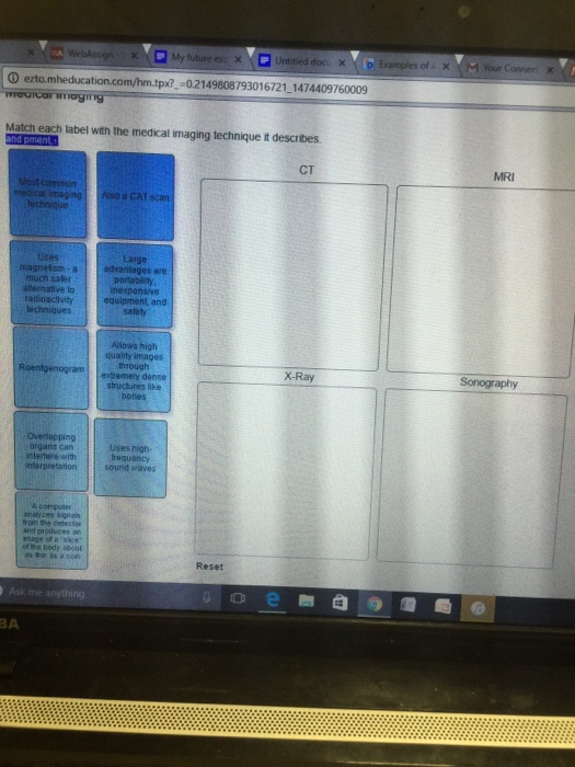
Solved Match Each Label With The Medical Imaging Technique Chegg Com
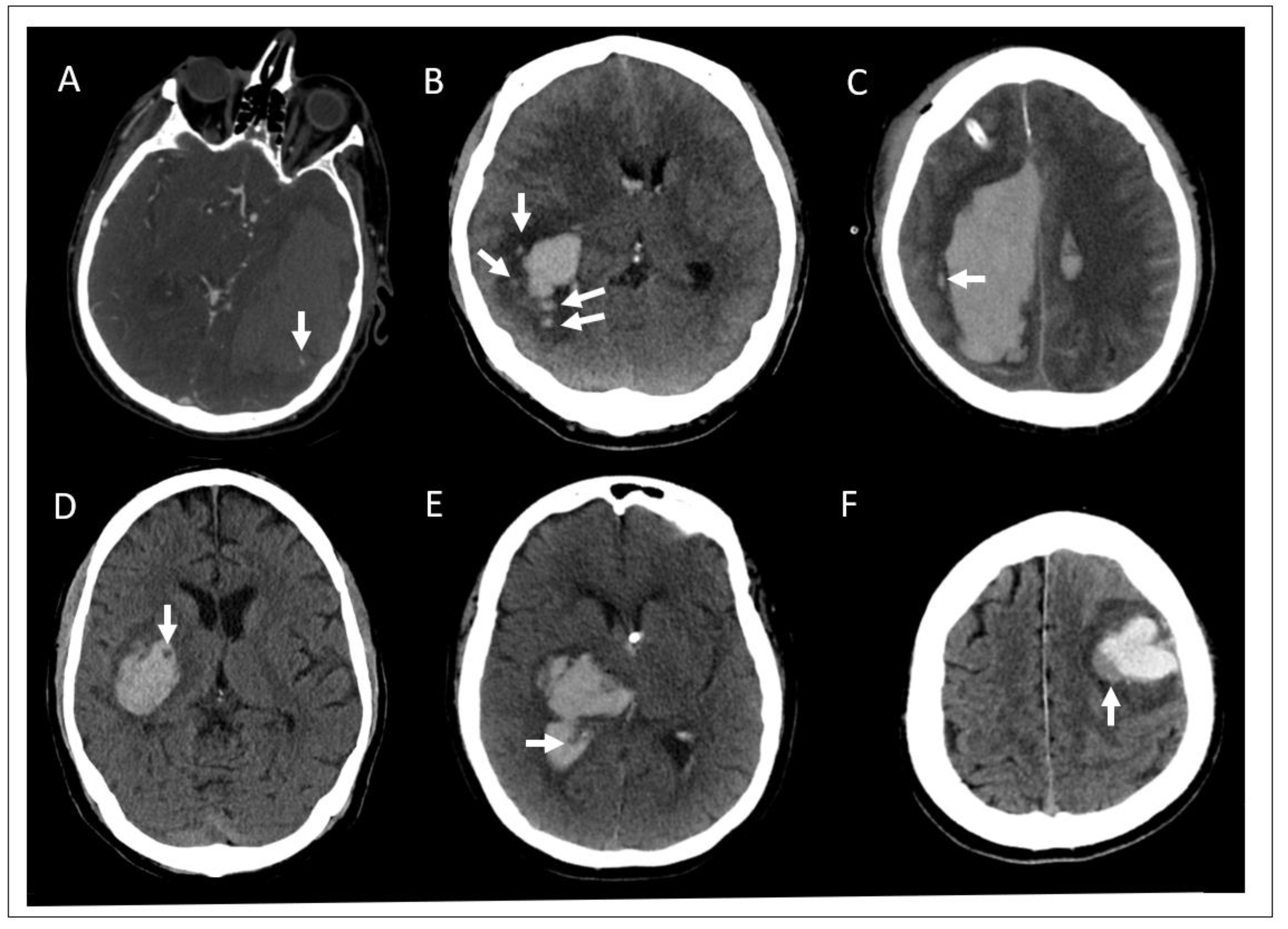
Diagnostics Free Full Text Minimally Invasive Intracerebral Hemorrhage Evacuation Techniques A Review Html
Medical Imaging Of Tissue Engineering And Regenerative Medicine Constructs Biomaterials Science Rsc Publishing Doi 10 1039 D0bm00705f
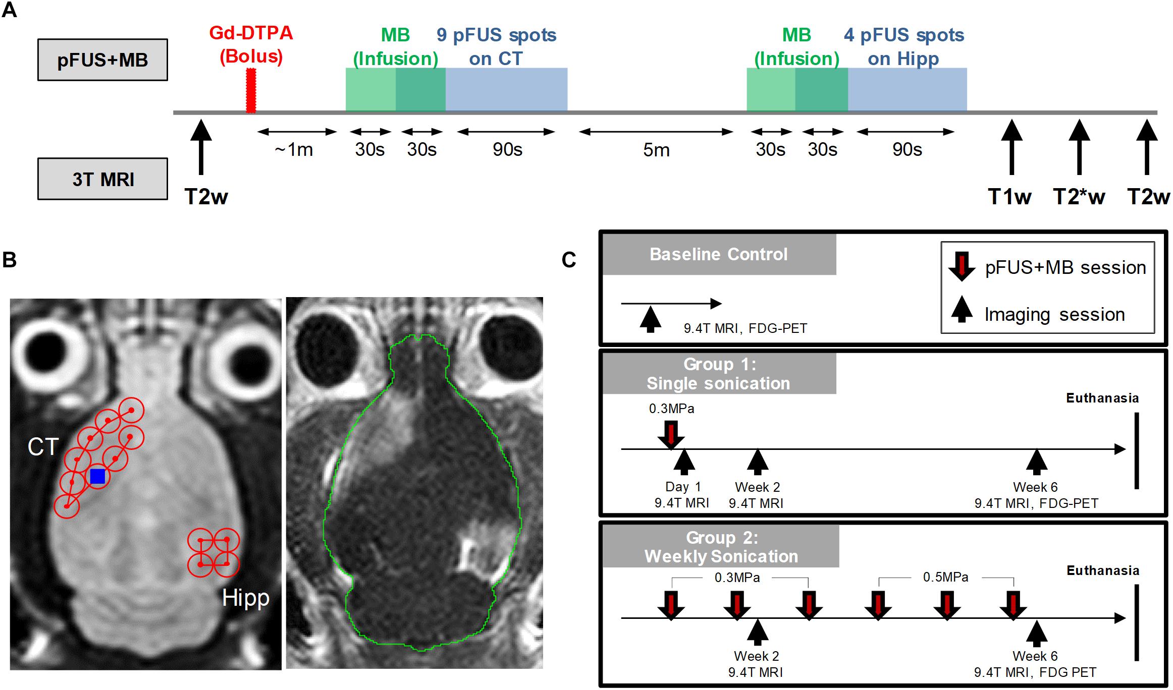
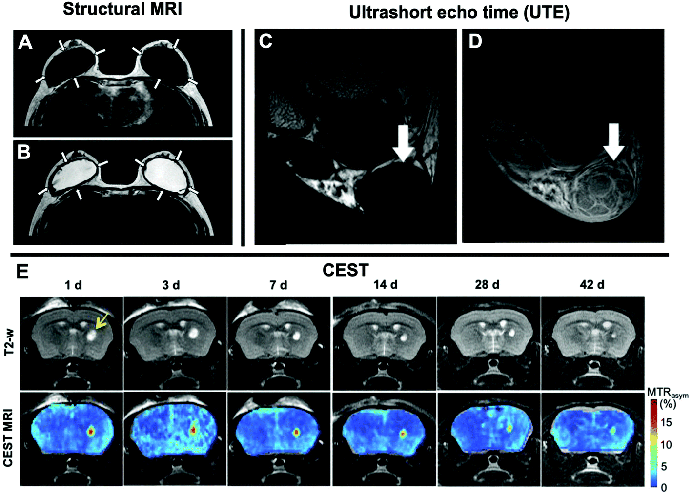
Frontiers Diffusion Tensor Imaging And Chemical Exchange Saturation Transfer Mri Evaluation On The Long Term Effects Of Pulsed Focused Ultrasound And Microbubbles Blood Brain Barrier Opening In The Rat Neuroscience
Diagnostic Pathways In Crohn S Disease Clinical Radiology

Arterial Spin Labelling Qualitative Assessment In Paediatric Patients With Mri Negative Epilepsy Clinical Radiology

A Review Of Medical Image Data Augmentation Techniques For Deep Learning Applications Chlap 2021 Journal Of Medical Imaging And Radiation Oncology Wiley Online Library

Personalising Sarcoma Care Using Quantitative Multimodality Imaging For Response Assessment Clinical Radiology
State Of The Art Techniques Using Pre Operative Brain Mri Scans For Survival Prediction Of Glioblastoma Multiforme Patients And Future Research Directions Springerlink
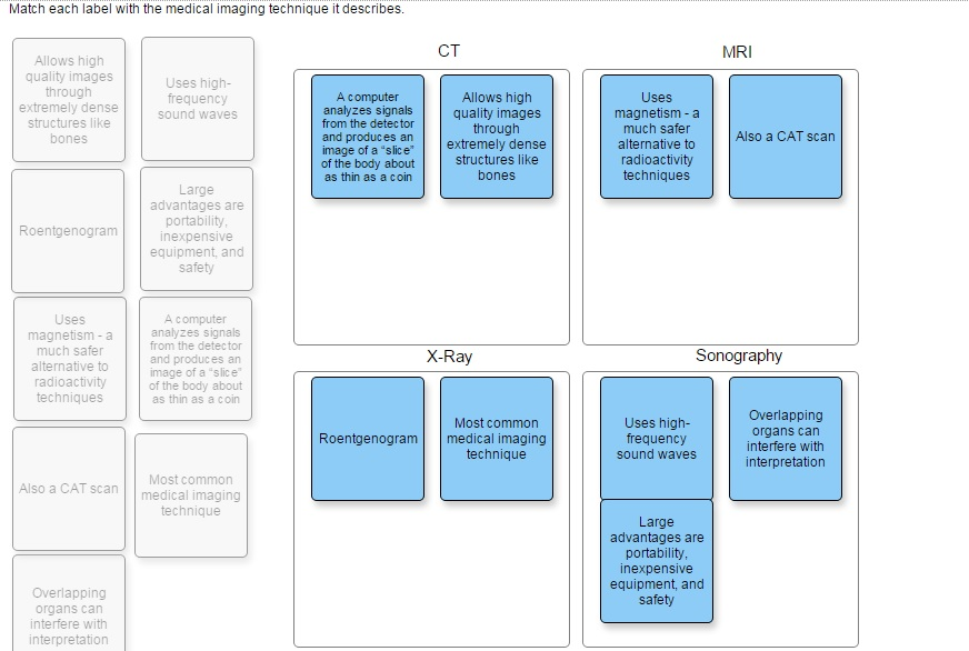
33 Match Each Label With The Medical Imaging Technique It Describes Label Design Ideas 2020
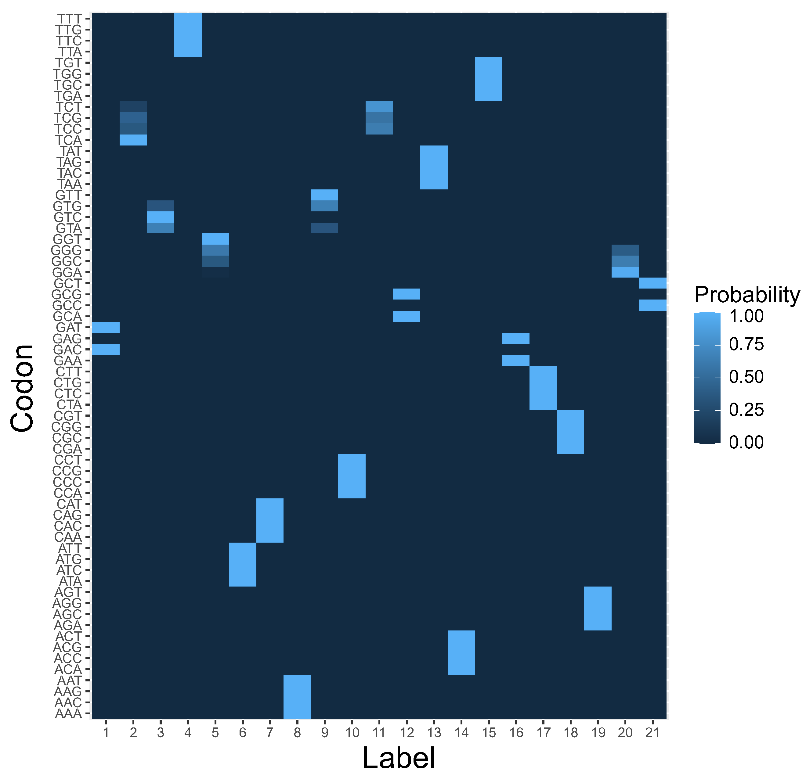
Ijms Free Full Text Model Of Genetic Code Structure Evolution Under Various Types Of Codon Reading Html


Comments
Post a Comment